Blogs
Avoiding Gross Mistakes in X-ray, CT or MRI Scanning Process
Images of Xray or CT scans or MRI scans can create confusion for radiologists. The right and left sides of the body are mistaken for one another as one side is mirror image of the other. When an image is electronically inverted, it is possible for laterality to be mistaken. As an example, the frontal view of the hand, whether on film or digitized, is normally taken with the palm of the hand against the cassette, resulting in an anteroposterior (AP) image.
The resulting image, however, can be viewed from either the front or the back. If viewed from the front or exposure side, the side from which the X-ray beam originally entered the body, it will appear to be a left hand. But if the same image is looked upon from the back-the opposite surface or nonexposure side, the viewer sees the mirror image, which appears to be a right hand.
Option1: Physical Marking:: Whenever a radiograph is made, it is important for the technitian to identify which side of the body appears on the image. This is usually achieved by placing a lead or a pen marker on the front of the CASSETTE (as shown in Pic01), corresponding to the side examined.
Option 2: Alternatively, a label is attached directly to the film after it is developed on a system, indicating which side is represented thereon. Since humans are occasionally fallible, however, this marking may be incorrect or omitted altogether (as shown in Pic03).
Hence, when the image is interpreted, there may be a question regarding which side of the body-right or left-actually appears on the image. This often occurs in busy radiology departments, especially in emergency departments (EDs). When an error is suspected in marking which side of the body has been examined there are two main possible outcomes.
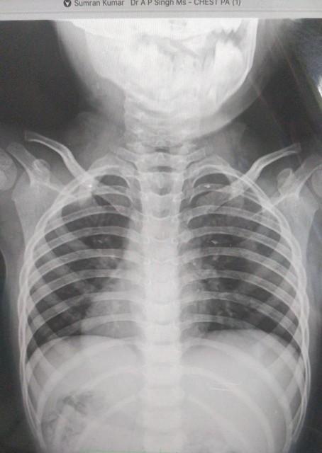
Radiographers/technitians are taught from day one in school to place anatomical markers within the primary beam of radiographs. We do so as a method of "best practice" to properly distinguish the patient's right from left on the radiographic image per legal requirements. Conditions like Dextrocardia (when the heart is positioned on the right instead of the left) and situs inversus (when all of the internal organs are on the opposite side compared to normal anatomy) as shown in Pic 02 exist which can easily be misinterpreted and would normally cause a technologist to inappropriately orient the image to appear similar to normal anatomy. But when radiographs misrepresent right from left, this presents a huge risk for medical errors.


When you subscribe to the blog, we will send you an e-mail when there are new updates on the site so you wouldn't miss them.
About the author
Latest Comments

Comments